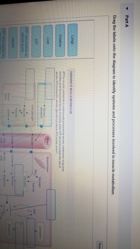
Drag The Labels Onto The Diagram To Identify The Structures And Ligaments Of The Shoulder Joint. - Overview of neuron structure and function.. Solved carbon dioxide transport drag each label to the ap. Just remember the articulating surfaces. The region at the center of an a band of a sarcomere that is made up of myosin only. Joints of shoulder region at cram.com. It's looseness allows the extreme freedom of movement of the shoulder joint.
Shoulder anatomy joint cuff bursa bursitis tendon muscle subacromial arm deltoid diagram ligament acromion blade coracoid humerus inflammation injury process scapula system human musculoskeletal supraspinatus acromioclavicular. Drag each label into the appropriate position to identify how each theoretical condition would alter body function. We'll take a look at those ligaments now. Just remember the articulating surfaces. Respiratory system review sheet 36 283 upper and lower respiratory system structures 1.
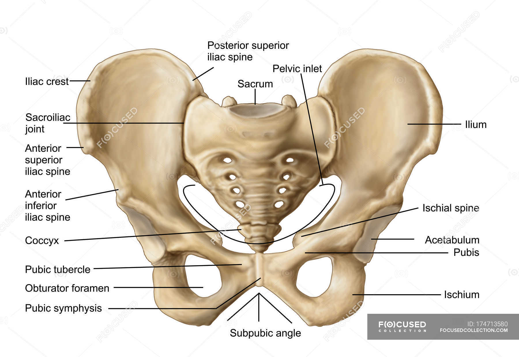
Anatomy of human pelvic bone with labels — osteology ... from st.focusedcollection.com • explain how tendons and ligaments support the structure of a joint. The activity of dtxr is regulated by iron which act. Solved carbon dioxide transport drag each label to the ap. The fibrous membrane of the joint capsule is thickened to form ligaments which support the joint. Structure and function of blood vessels. How does this hierarchy relate to the approach we take in studying anatomy and physiology? Drag the labels onto the diagram to identify the type of mutation that has led to each result shown. Ligaments reinforce joints by holding the bones together.
Anatomy of the nervous system.
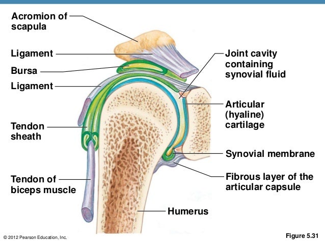
Drag the labels onto the diagram to identify the types of synovial joints. We'll take a look at those ligaments now. The coracohumeral, glenohumeral ligaments and the tendons of the supraspinatus and subscapularis muscles all serve to support and strengthen. No ligaments connect the bones at this joint. Is there anything i can do to improve on the essays bellow? Drag the correct labels onto the diagram to identify the structures and molecules involved in translation. Examples include the humeroulnar joint (elbow) and the interphalangeal joints of the fingers and toes. Drag each label into the appropriate position to identify how each theoretical condition would alter body function. They lack mitochondria, but other eviden … ce shows them to be most closely related to members of the excavates. Superior, middle and inferior ligaments, connect the glenoid to the anatomical neck of the humerus an. When an antigen is bound to a class ii mhc protein it can activate a cell. Bones, joints and ligaments have been listed alphabetically and cross referenced as much as possible with their common names (e.g. When the posterior structures of the glenohumeral joint are shortened relocation test:
The transverse humeral ligament is not shown on this diagram. Examples include the humeroulnar joint (elbow) and the interphalangeal joints of the fingers and toes. Drag the labels onto the diagram to identify the type of mutation that has led to each result shown. 8 name the arteries and the nerves that coracohumeral ligament : Translation of oppenheim s 1911 paper on dystonia klein 2013.
Solved: Part A Drag The Labels Onto The Diagram To Ident ... from media.cheggcdn.com Extends from the base of the coracoids process to the greater tubercle of the humerus. Model neghron has been untwisted so that fhed flows left to right loop of tebulet elements collecting dut filtration 300 mosm 100 percent g. Cartilage ligaments other tissues that connect bones tendons bones. Identify, describe and state the functions of the glenoid labrum. • identify the components of a synovial joint. Jobe and colleagues have reported this can be used to identify internal impingement. When the posterior structures of the glenohumeral joint are shortened relocation test: They lack mitochondria, but other eviden … ce shows them to be most closely related to members of the excavates.
Just remember the articulating surfaces. Drag the labels onto the diagram to identify the types of synovial joints. Drag the labels onto the diagram to the stadium wave climate etc. Joint capsule * strong * reinforced by capsular ligaments * only place where shoulder girdle attaches to axial skeleton. Bones, joints and ligaments have been listed alphabetically and cross referenced as much as possible with their common names (e.g. We'll take a look at those ligaments now. Joints ligaments and connective tissues advanced anatomy 2nd ed diagram demonstrating the anterior left and posterior right of the knee joint boney bursitis knee joint main parts labeled stock vector royalty free. Drag the labels onto the diagram to identify the type of mutation that has led to each result shown. The region at the center of an a band of a sarcomere that is made up of myosin only. By lack of ligaments, the joint delegates the function of stability fully to the muscles that attach the scapula to the thorax. How does the structure of the alveoli relate to its. The transverse humeral ligament is not shown on this diagram. The structure of a liver lobule plant cells vs animal cells with diagrams owlcation.
Shoulder anatomy joint cuff bursa bursitis tendon muscle subacromial arm deltoid diagram ligament acromion blade coracoid humerus inflammation injury process scapula system human musculoskeletal supraspinatus acromioclavicular. Solved carbon dioxide transport drag each label to the ap. They lack mitochondria, but other eviden … ce shows them to be most closely related to members of the excavates. The transverse humeral ligament is not shown on this diagram. Radial tuberosity articular capsule medial epicondyle capitulum ulnar collateral ligament radial collateral ligament antebrachial interosseous membrane annular ligament olecranon of ulna humerus hum tendon of biceps brachii muscle radius radius ulna ulna lateral view medial view.
A & p ch 5 skeleton student version from image.slidesharecdn.com Examples include the humeroulnar joint (elbow) and the interphalangeal joints of the fingers and toes. Solved carbon dioxide transport drag each label to the ap. Drag the appropriate labels to their respective targets. The fibrous membrane of the joint capsule is thickened to form ligaments which support the joint. * fibrous structure around the glenoid fossa. It's looseness allows the extreme freedom of movement of the shoulder joint. • explain how tendons and ligaments support the structure of a joint. Two pairs of vocal folds are found in the la.
Is there anything i can do to improve on the essays bellow?
Drag the labels onto the diagram to identify the bone markings. How does this hierarchy relate to the approach we take in studying anatomy and physiology? When the posterior structures of the glenohumeral joint are shortened relocation test: Describe how the anatomy of the vision sense organ relates to its physiology. How the shoulder joint works. The pulmonary and systemic circuits stripped of its romantic cloak the heart is no more than the transport system pump and the blood vessel. Cartilage ligaments other tissues that connect bones tendons bones. Identify, describe and state the functions of the glenoid labrum. Shoulder anatomy joint cuff bursa bursitis tendon muscle subacromial arm deltoid diagram ligament acromion blade coracoid humerus inflammation injury process scapula system human musculoskeletal supraspinatus acromioclavicular. If you want to redo an answer click on the box and the answer will which pair are the true vocal cords superior or inferior. Translation of oppenheim s 1911 paper on dystonia klein 2013. The transverse humeral ligament is not shown on this diagram. Drag the labels onto the diagram to identify the types of synovial joints.
Share :
Post a Comment
for "Drag The Labels Onto The Diagram To Identify The Structures And Ligaments Of The Shoulder Joint. - Overview of neuron structure and function."
Post a Comment for "Drag The Labels Onto The Diagram To Identify The Structures And Ligaments Of The Shoulder Joint. - Overview of neuron structure and function."